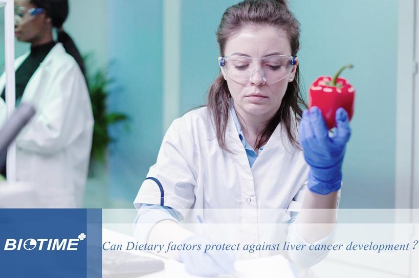
immunofluorescence reagents
  • Blockbuster!Xiamen Biotime has been awarded MDSAP certification
    Blockbuster!Xiamen Biotime has been awarded MDSAP certification July 22, 2022
    Recently, Xiamen Biotime Biotechnology Co., Ltd. completed the Medical Device Single Audit Program (MDSAP) and obtained the MDSAP certification issued by the international certification body SGS MDSAP Certification Program It is co-sponsored by members of the International Medical Apparatus Regulators Forum (IMDRF) and recognized and joined by regulators in the United States (FDA), Australia (TGA), Brazil (ANVISA), Canada (HC), and Japan (MHLW). A new audit procedure allows medical apparatus manufacturers to undergo only one quality management system audit to meet the standards and regulatory requirements of the above five countries. The audit work is carried out by an audit body authorized by the regulatory agencies of the five countries. As a high-tech enterprise specializing in the research and development, production and sales of in vitro diagnostic reagents and instruments, Xiamen Biotime Biotechnology Co., Ltd. always adheres to the quality policy of 'product-oriented, innovation-oriented, management-oriented, and service-oriented', and strictly controls product quality management. Obtaining the MDSAP certification will further bring advantages and convenience to Xiamen Biotime's products to obtain overseas market access qualifications, and accelerate the pace of the company's global development strategy. In the future, Xiamen Biotime Biotechnology Co., Ltd. will continue to help the global epidemic prevention and control work, be committed to providing high-quality in vitro diagnostic products to more countries and regions around the world, and contribute to the development of the global biomedical industry.
    View More
  • What kinds of POCT HbA1c devices have been available in the market?
    What kinds of POCT HbA1c devices have been available in the market? June 21, 2021
    Diabetes mellitus(DM)is a chronic disease caused by deficiency or diminished effectiveness of endogenous insulin. It is characterised by hyperglycaemia, deranged metabolism and sequelae predominantly affecting the vasculature. The most common types include type 1 and type 2 DM. DM represents a major health problem of the 21st century, causing severe long-term damage to the cardiovascular and nervous system as well as eyes and kidneys. The World Health Organization (WHO) estimated 1.6 million deaths each year to be directly caused by diabetes [1]. Most cases (90%) of DM are type 2, which arises from defects in insulin action leading to insulin resistance, often combined with defects in insulin secretion. Circulatory insulin levels therefore may be normal or raised, but it cannot be used effectively. This subtype is predominant in middle-aged overweight patients with a sedentary lifestyle. In 2011 the WHO advocated the use of HbA1c for the diagnosis of type 2 DM and in 2012 UK guidance followed suit [2, 3]. The term HbA1c refers to glycated haemoglobin. It develops when haemoglobin, a protein within red blood cells that carries oxygen throughout your body, joins with glucose in the blood, becoming “glycated”. HbA1c test tells you your average level of blood sugar over the past 2 to 3 months. Most POCT devices for HbA1c use a drop of capillary or venous whole blood. Following application to the test cartridge, the sample is analysed within a few minutes using methods based on either differences in structure or charge of the glycated vs non-glycated haemoglobin. Ion-exchange chromatography: Haemoglobin species (HbA1c and HbA0) are separated based on the difference in isoelectric point, by employing differences in ionic interactions between the haemoglobin in the blood sample and the cation exchange groups on the column resin surface. HPLC is based on ion-exchange chromatography technology, which is a gold standard in detecting HbA1c. Immunoassay: The immunoassay method uses antibodies which bind to the N-terminal glycated tetrapeptide or hexapeptide group of the HbA1c, forming immunocomplexes which can be detected and measured using a turbidimeter or a nephelometer. Affinity chromatography: Affinity chromatography is a separation technique based on structural differences between glycated vs non-glycated haemoglobin which utilises m-aminophenylboronic acid and its specific interactions with the glucose adduct of glycated haemoglobin. Enzymatic assay: Enzymatic quantification of HbA1c is based on cleavage of the beta chain of haemoglobin by specific proteases to liberate peptides, which then further react to produce a measurable signal (4). HbA1c in diagnosis HbA1c can indicate people with prediabetes or diabetes as follows: How often do we need to take a HbA1c test? Everyone with diabetes mellitus should be offered a HbA1c test at least once a year. Some people may have an HbA1c test more often. T...
    View More
  • [Biotime announcement] - Biotime infection marker interleukin 6 (IL-6) is officially launched!
    [Biotime announcement] - Biotime infection marker interleukin 6 (IL-6) is officially launched! April 22, 2021
    Interleukin is a type of cytokine produced by and acting on a variety of cells. Interleukin 6 (Interleukin 6, abbreviated IL-6) is an important member of the cytokine network, and it has been widely used in many clinical fields. PCT has been widely recognized clinically in differential diagnosis of bacterial infection, risk stratification of sepsis and guidance of antibiotic use. IL-6 is an important cytokine expressed in the initial response of the innate immune system to injury and infection, and plays an important role in the early diagnosis of acute infection. IL-6 combined with PCT has complementary advantages, improves detection sensitivity and specificity, and better facilitates the management of the whole course of sepsis. IL-6 is also one of the important monitoring indicators during the diagnosis and treatment of COVID-19. IL-6 is used for early warning of sepsis After the occurrence of inflammation, IL-6 is the first inflammatory marker. IL-6 will be involved in the occurrence and development of many diseases, and its blood level is closely related to inflammation, viral infection and autoimmune diseases, and its changes are earlier than CRP and PCT. Studies have shown that IL-6 increases rapidly after bacterial infection, PCT increases after 2h, and CRP increases rapidly after 6h [1]. So IL-6 can be used for early warning of sepsis. (Kinetic changes of inflammatory markers in vivo after endotoxin stimulation) IL-6 is used to evaluate the prognosis of sepsis Studies have shown that the high IL-6 concentration during the treatment of sepsis indicates the poor prognosis of patients and the higher mortality rate at the later stage of treatment. The expert consensus on the interpretation of the clinical significance of infection-related biomarkers mentioned that IL-6> at 1000 pg/mL suggests a poor prognosis. Therefore, the detection of IL-6 is also of great significance for the prognosis assessment of sepsis. Application of IL-6 in COVID-19 IL-6 is an important pathway that induces cytokine storm. Cytokine storm is a kind of transitional immune response, and the uncontrolled release of inflammatory factors eventually leads to organ damage and even functional failure. From a large number of research data, it is generally believed that cytokine storm is a key factor in the deterioration of COVID-19 patients. Therefore, IL-6 is also an important monitoring indicator in the diagnosis and treatment of COVID-19, which can be used for early warning of severe/critical COVID-19 and can also help guide the clinical selection of appropriate treatment strategies. a) Key laboratory indicators for COVID-19 patient assessment b) IL-6 levels in 99 confirmed COVID-19 patients Figure (a) In a retrospective multicentric study of 150 confirmed COVID-19 cases, IL-6 in the deceased patients was significantly higher than that in other cases [2]. Figure (b) In a study of 99 patients published in the January 29 Lancet, IL-6 was significantly elevated in more th...
    View More
  • What you should know about D-Dimer?
    What you should know about D-Dimer? June 16, 2021
    D-Dimer is one of the biomarkers with high attention on the market in recent years, which is used to indicate thrombosis and thrombolytic activity in human body. Let’s first talk about its clinical application. 1. Venous Thromboembolism (VTE) the level of D-Dimer is significantly increased in VTE patients, however, some diseases such as cardiovascular disease, surgery, tumor, infection, and tissue necrosis also have a certain impact on blood clotting, which cause D-Dimer to increase. Therefore, the sensitivity of D-Dimer is 97%, but the specificity is around 61%-64%[1]. So D-Dimer can not be used to diagnose VTE alone. The practical significance of D-Dimer is that a negative result can exclude VTE. 2. Deep vein thrombosis (DVT) a blood clot that's deep inside a vein. These clots usually affect the lower legs, but they can also happen in other parts of the body. 3. Pulmonary embolism (PE) a blockage in an artery in the lungs. It usually happens when a blood clot in another part of the body breaks loose and travels to the lungs. DVT clots are a common cause of PE. 4. Disseminated intravascular coagulation (DIC) DIC is a syndrome characterized by the systemic activation of blood coagulation, which generates intravascular fibrin, leading to thrombosis of small and medium-sized vessels, and eventually organ dysfunction. DIC may occur as a complication of infections, solid cancers, hematologic malignancies, obstetric diseases, trauma, aneurysm, and liver diseases, etc. International Society on Thrombosis and Haemostasis(ISTH) suggested that a reduced fibrinogen level is valuable for the diagnosis of DIC, but is not observed in most DIC patients. The elevated fiber-related markers (FRMs), such as fibrinogen and fibrin degradation products, D-dimers, and soluble fibrin (SF), reflect thrombin formation[3]. 5. D-Dimer and COVID-19 Correlations exist between COVID-19 infection, severe elevation of D-dimer levels, and increase in the rate of complications and composite end-point. The appropriateness of early and continuous D-dimer monitoring and labeled anticoagulation as management tools for COVID-19 disease deserves accurate investigation, to prevent complications and reduce interventions[4]. Chinese Clinical Guidance for COIVD-19 Pneumonia Diagnosis and Treatment(7th edition) recommended that D-Dimer can be used for the diagnosis of severe COIVD-19 patients. What is the best test method in detecting D-Dimer? ① Quantitative Test ② High specificity and negative predictive value ③ Good repeatability ④ The coefficient of variation is low when the result is at the critical level ⑤ Rapid and convenient What could cause D-Dimer false negative? ① The volume of the thrombus is small or the distal small thrombus ② Fibrinolytic activity is decreased ③ Unreasonable design of reference interval and threshold ④ There are problems with sample preparation In summary, a rapid, convenient, and quantitative D-Dimer test with high sensitivity has been widely u...
    View More
  • Biotime attended medical trade fair CACLP 2021
    Biotime attended medical trade fair CACLP 2021 March 31, 2021
    Established in 1991, CACLP Expo now is the largest, the most influential and professional trade show in China In Vitro Diagnostics (IVD) and clinical laboratory industry. Biotime, a leading manufacturer of POCT, attended China Association of Clinical Laboratory Practice (CACLP) Expo 2021 from March 28th to 30th at Chongqing International Expo Center. During this CACLP, Biotime exhibited a full line of Point of Care Testing (POCT) reagents and released 3 automated analyzers, named HLC-100 Fully Automated HbA1c Analyzer, FLI-600 FIA Analyzer and FLI-4000 Fully Automated FIA Analyzer. At the same time, Biotime brought a wonderful academic satellite symposium. Cardiology expert Wang Bin was invited to introduce the chest pain center and related cardiac markers. Biotime’s new innovations attracted lots of medical experts and distributors' attention to stop by and communicate with each other. We are so grateful for visitors' support and wish to meet you next year!
    View More
  • How Productive is the D-Dimer for Chest Pains and Covid-19?
    How Productive is the D-Dimer for Chest Pains and Covid-19? March 02, 2022
    How Productive is the D-Dimer for Chest Pains and Covid-19?   D-dimers are cleavage products of fibrin that occur during plasmin-mediated fibrinolysis of blood clots. In the emergency department, D-dimer tests are broadly used as an excellent non-invasive triage biomarker in patients, which measurement represents a valuable and cost-effective tool in the differential diagnosis of acute chest pain including the main life-threatening entities: acute coronary syndrome, pulmonary embolism, and acute aortic syndrome. Whereas the diagnostic and prognostic values of D-dimer testing in acute coronary syndrome is of less priority, increases of D-dimers are frequently found in venous thromboembolism and acute aortic syndromes, especially acute aortic dissection.   Laboratory evaluation   D-dimer levels can be assessed by microplate enzyme-linked immunosorbent assay, enzyme-linked immunofluorescence assay, whole-blood cell agglutination, or latex agglutination tests. The sensitivity, specificity, and negative predictive value of D-dimer tests depend on the kind of used test, the cutoff value, and the kind of assumed disease. Cutoff levels are influenced by the kind of test used and may differ between different laboratories. D-dimer levels are associated with the amount of clotted blood. Highest levels are reached in massive venous thromboembolism and after cardiovascular arrest.   Influence of D-Dimers on ACS   Acute myocardial ischemia is caused by thrombotic occlusion of coronary arteries, implicating that D-dimer levels should be raised in focal coronary thrombosis. Whereas troponin is a highly sensitive and specific parameter for myocardial injury, elevation is measurable not until 3-4 hours after onset of symptoms. By contrast, D-dimer propose earlier rise than common markers of cardiac injury. It has been demonstrated that increased D-dimer levels may serve as an independent diagnostic marker for myocardial infarction with an increase in diagnostic sensitivity of the electrocardiogram and clinical history plus D-dimer.   Value of D-Dimer Measurement in PE   D-dimers have been shown to be highly sensitive in venous thromboembolism. Owing to the fact that deep vein thrombosis and pulmonary embolism often occur at the same time and up to 50% of patients with deep vein thrombosis have clinically inapparent pulmonary embolism, D-dimer levels can be used similarly in patients with deep vein thrombosis and pulmonary embolism. Therefore, the D-dimer test as a fast and cost-effective method is recommended in suspected pulmonary embolism.   D-Dimers in Aortic Dissection and other Aortic Syndromes   The term acute aortic syndrome includes different conditions ranging from an intramural hematoma to typical aortic dissection, together characterized by a disruption of the structural integrity of the aortic wall with subsequent initiation of coagulation. Apart from clinical presentation, imaging methods, and other biomark...
    View More
  • Biotime received The Medical Devices Excellence Awards
    Biotime received The Medical Devices Excellence Awards July 02, 2021
    Great News On 17th June, Biotime proudly received The Medical Devices Excellence Awards for its BIOT-YG-I FIA Analyser, a recognition for Chinese organizations that managed to produce the greatest achievements and innovations in the Chinese medical devices industry. All of our efforts at Biotime are part of our uncompromising commitment to generating superior health-related products, addressing new and growing patient needs. In light of this, Biotime will continue to push forward with self-dependent innovations, launching new solutions in multiple sectors and ultimately making contribution to the totality of progress in improving the health and wellbeing of humankind. Winner of the Medical Devices Excellence Awards - BIOT-YG-I FIA Analyser
    View More
  • The development of the boronate affinity chromatography and Biotime’s success
    The development of the boronate affinity chromatography and Biotime’s success March 14, 2022
    Boronate affinity chromatography (BAC) is a unique means for selective separation and enrichment of cis-diol-containing compounds. Cis-diol-containing biomolecules are an important class of compounds, including glycoproteins, glycopeptides, ribonucleosides, ribonucleotides, saccharides, and catecholamines. These biomolecules play essential roles in many life-related processes. Because cis-diol-containing biomolecules are important target molecules in current research frontiers such as proteomics, metabolomics, and glycomics, BAC and boronate affinity materials have gained rapid development and found increasing applications in recent decades. BAC is a unique mode of affinity chromatography, in which a boronic acid is used as the ligand for the selective isolation and enrichment of cis diol-containing compounds. The retention mechanism mainly relies on the pH-controlled reversible covalent interactions between cis-diol groups and the boronic acid ligand. As compared to other affinity chromatographic techniques, BAC exhibits several significant features, including broad-spectrum selectivity, reversible covalent binding, pH-controlled binding/release, and fast association/desorption kinetics. Owing to these merits, BAC is of great value in a variety of fields such as affinity separation, proteomic analysis, and metabolomics analysis.   HISTORICAL DEVELOPMENT The history of BAC can be simply divided into three different periods: early development period before 1970, approach-forming period 1970–2005, and new development period since 2006.   PRINCIPLE AND BINDING PH BAC principle relies on the reversible covalent reaction between cis-diol-containing compounds and boronic acid ligands. Figure 1 shows a general formula for the interaction between boronic acid and a cis diol-containing compound. When the surrounding pH is greater than the pKa value of the boronic acid, hydrolysis of the boronic acid occurs, resulting in a hybridization status change from trigonal coplanar shape to tetragonal boronate anion (from sp2 to sp3). The obtained tetragonal boronate anion can react with cis-diols and form five or six-membered cyclic esters. When the pH of the surrounding solution is switched to acidic, the boronic acid-cis-diol complex dissociates, because the binding strength between boronic acids in trigonal form and cis diol-containing compounds is very weak. Owing to the pH-controlled reversible covalent reaction, elution of captured analytes in BAC is very simple, just needing an acidic solution as the eluting buffer. Alternatively, the release of the captured analytes by the boronic acid ligands can be realized through adding excessive amounts of competing for cis-diol-containing molecules such as sorbitol into the loading buffer.   BORONATE AFFINITY CHROMATOGRAPHY Figure 1 Schematic diagram showing the interaction between boronic acids and cis-diol-containing compounds. INTERACTION MECHANISM AND SELECTIVITY MANIPULATION Selectivity is an esse...
    View More
First 1 ... 5 6 7 8 Last
[  A total of  8  pages]
Leave A Message

Leave A Message

    Please provide us with the information below, and we'll contact you as soon as possible.

Home

products

about

contact