immunofluorescence reagents
  • IL6-A ROBUST PREDICTOR OF SEVERE COVID19 ILLNESS
    IL6-A ROBUST PREDICTOR OF SEVERE COVID19 ILLNESS July 29, 2021
    WHAT IS IL-6 IL-6 is an immune protein promptly and transiently produced in response to tissue damage and infections. On the other hand, IL-6 is also a pyrogen and is responsible for fever in autoimmune, infectious, or non-infectious diseases. IL-6 is produced in the body, wherever there is inflammation, either acute or chronic. This includes situations such as trauma, burns, cancers, and infection. (Thomas, 2021)   RELATIONSHIP BETWEEN IL-6 AND COVID-19 During the pandemic, scientists have explored the role of IL-6 as a predictor for patients with severe coronavirus disease.   Based on the finding that infection by the severe acute respiratory syndrome-associated coronavirus induces a dose-dependent production of IL-6 from bronchial epithelial cells, a study involving nearly 11,000 patients discovered that treating hospitalized COVID-19 patients with drugs that block the effects of interleukin-6 reduces the risk of death and the need for mechanical ventilation. (Henderson, 2021)   According to the study, in hospitalized patients, administering the interleukin-6 antagonists (such as tocilizumab and sarilumab) in addition to corticosteroids reduces the risk of death by 17%, compared to the use of corticosteroids alone. In patients not on mechanical ventilation, the risk of mechanical ventilation or death is reduced by 21%, compared to the use of corticosteroids alone. (Henderson, 2021)   Some also conducted data analysis of COVID-19 cases at long-term care facilities in Slovakia Patients with hypoxemia had significantly higher concentrations of IL-6, C-reactive protein, procalcitonin, fibrinogen, total bilirubin, aspartate aminotransferase and alanine aminotransferase at initial screening among which IL-6 was identified as the most robust predictor of hypoxemia. (Sabaka et al., 2021)   Given the current situation of global vaccine inequity, people in the lowest income countries will be the ones most at risk of severe and critical COVID-19. Therefore, further work is necessary to clarify the exact role of IL-6 during coronavirus infections and the potential role of this cytokine to be used as a biomarker of viral virulence. Additional work exploring the potential therapeutic use of blocking IL-6 or different products affected by its activity might provide insight into controlling persistent COVID-19 infections. (Velazquez-Salinas et al., 2019)   BIOTIME IL-6 Biotime IL-6 is an in vitro diagnostic test for the quantitative determination of IL 6 in human serum or plasma. As researchers have pointed out a portion of severely ill COVID-19 patients experience rapid deterioration and need for mechanical ventilation due to development of ARDS. Being an unconventional way to address the issue, Biotime IL-6 can also be used to assist in identifying severe inflammatory response in patients with confirmed COVID 19 illness who need mechanical ventilation in conjunction with clinical findings.     References Thomas, L. (2...
    View More
  • What is an IgE test?
    What is an IgE test? July 12, 2021
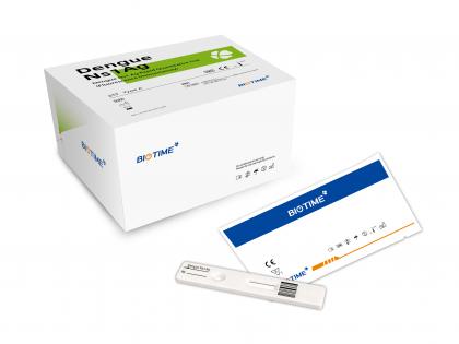
    Immunoglobulin E (IgE) is an antibody produced by the body's immune system in response to a perceived threat. It is one of the five types of immunoglobulins (A, G, M, D, and E) and is usually found in very small amounts in the blood. The total IgE test can be used to help screen and detect allergic diseases. It measures the total amount of immunoglobulin E in the blood. Immunoglobulin is a protein that plays a key role in the human immune system. They are produced by specific immune cells called plasma cells. Immunoglobulins are produced against bacteria, viruses, and other microorganisms and substances that are recognized as "non-self" or provide harmful antigens to the immune system. Elevated concentrations of total immunoglobulin E (IgE) can be found in a variety of clinical diseases, including allergic diseases, certain primary immune deficiencies, infections, inflammatory diseases, and malignant tumors, and to a lesser extent, are related to the immunity of parasites is related. Occasionally, an IgE test may be needed to help diagnose a very rare genetic disease called high immunoglobulin E syndrome (Job syndrome). People with this disease usually have significantly higher IgE levels than normal, and may have eczema, recurrent sinus and lung infections, bone defects, and severe skin infections. A large increase in IgE concentration may indicate that a person has inherited this condition. When a person experiences periodic or persistent symptoms due to an allergic reaction, especially when the underlying allergen is unknown, a total IgE test may be required. Symptoms may include those that indicate involvement of the skin, respiratory system, and/or digestive system, such as: -Periodic or persistent itching -Urticaria -Itchy eyes -Eczema -Nausea, vomiting, persistent diarrhea -Sneezing, coughing, congestion -Difficulty breathing -Asthma symptoms: wheezing, difficulty breathing, coughing, chest tightness Biotime IgE rapid quantitative test kit is in great demand right now. It allows use of different sample material: whole blood, plasma, and serum with results conveniently available, making the process a lot less cumbersome and genuinely helping screen and detect a variety of clinical diseases. Reference 1. Homburger HA: Allergic diseases. In: Clinical Diagnosis and Management by Laboratory Methods. 21st ed. WB Saunders Company. 2007;961-971 2. Martins TB, Bandhauer ME, Bunker AM, Roberts WL, Hill HR: New childhood and adult reference intervals for total IgE. J Allergy Clin Immunol. 2014 Feb;133(2):589-591 3. Bernstein IL, Li JT, Bernstein DI, et al: Allergy diagnostic testing: An updated practice parameter. Ann Allergy Asthma Immunol. 2008 Mar;100(3 Suppl 3):S1-148 4. Ansotegui IJ, Melioli G, Canonica GW, et al: IgE allergy diagnostics and other relevant tests in allergy, a World Allergy Organization position paper. World Allergy Organ J. 2020 Feb;13(2):100080. doi: 10.1016/j.waojou.2019.100080
    View More
  • COVID DELTA VARIANT: WHAT WE KNOW
    COVID DELTA VARIANT: WHAT WE KNOW July 09, 2021
    More than 18 month since the outbreak, COVID 19 is still raging in much of the world. The novel coronavirus has mutated into strains such as the Delta variant which was first detected in India and accounted for 99% of Covid-19 cases in the UK by mid-Jun, according to Public Health England. (The Guardian, 2021) Delta strain is a highly contagious and possibly more severe SARS-CoV-2 virus strain. Delta is spreading 50% faster than Alpha, which was 50% more contagious than the original strain of SARS-CoV-2—making the new variant 75% more contagious than the original. (YaleMedicine, 2021) And like we’re seeing now, Delta is outcompeting everything else and becoming the dominant strain. The symptoms of the Delta variant appear to differ from traditional COVID symptoms, According to data from the United Kingdom, the most common COVID symptoms may have changed from those we traditionally associated with the virus. Those include: Put aside fever and cough which have always been common COVID symptoms, and headache and sore throat which have traditionally presented for some people, a runny nose is now being reported significantly more frequently. Meanwhile, loss of smell, which was originally quite common, now ranks ninth. (Herrero, 2021) Although it's still not clear whether it it's more dangerous, scientists are becoming more confident the Delta variant represents a more transmissible SARS-CoV-2 strain. (Fox, 2021) What’s even more concerning, is that the Delta variant that has caused devastation in countries like India and the UK has now mutated to produce another variant called Delta Plus. (Joi, 2021) While vaccines are rolling out, not all of us are able to receive them as soon as we'd like. And because not all of us are yet fully protected, get tested if you have any symptoms, even if it’s “just a sniffle”, to protect yourself with some added peace of mind as well as others in your community. Through the course of the pandemic, Biotime’s consolidated its commitment to bringing accessible COVID-19 rapid self-testing to the masses. Hence a wide range of covid 19 test kits has been rolled out and made available both nationwide and worldwide. In particular, the BIOTIME SARS-CoV-2 IgG/IgM Rapid Qualitative Test is a lateral flow immunoassay intended for qualitative detection and differentiation of IgM and IgG antibodies to SARS-CoV-2 in human serum, plasma (potassium EDTA), and potassium EDTA venous whole blood. Our rapid antigen covid test kit is also qualified to detect both asymptomatic and symptomatic COVID-19, helping society collectively return to daily life. In addition to that, some scientific researches have purposefully assessed the use of D-dimer, PCT, CRP, IL-6, Ferritin as biomarkers for disease severity and mortality in COVID-19 patients. And the results are rather convincing to suggest they can provide important information on the prognosis of the novel corona virus infection. Below is Biotime’s product specification. Shop now on our websit...
    View More
  • Product Recommendations | NGAL- Early Marker of Acute Kidney Injury
    Product Recommendations | NGAL- Early Marker of Acute Kidney Injury July 05, 2021
    What is NGAL? The full name of NGAL is human lipocalin 2 (Ln2) or siderocalin. It belongs to the lipocalin family and is the earliest proposed marker of acute kidney injury (AKI).   What is the clinical significance of NGAL? 1. NGAL is a marker of acute kidney injury (AKI) When AKI occurs, NGAL rises rapidly in a very short period of time, and the most obvious is 2h. Serum creatinine (sCr), urinary enzymes, etc. often increase significantly after 24 to 72 hours. At this time, the glomerular filtration rate (GFR) has decreased significantly, and NGAL can change before GFR decreases, so it is suitable for early diagnosis of AKI. At the same time, the increase in NGAL concentration is closely related to the duration of AKI, the severity of kidney injury (RIFLE classification), and the treatment effect and prognosis. 2、Can predict the progression of chronic kidney disease (CKD), especially diabetic nephropathy Studies have found that NGAL may be involved in the occurrence and development of diabetes as an inflammatory factor. In diabetic nephropathy, renal tubule damage may be earlier than glomerulus. As a marker of early renal tubular damage, NGAL can reflect renal function damage in diabetic patients (especially type 2 diabetes) before urinary microalbumin (mALB). 3、NGAL also has a certain relationship with tumors Data from different laboratories indicate that the concentration level of NGAL is related to a variety of tumors. It promotes cell proliferation by uptake of iron ions and helps tumor cell growth and infiltration. It is related to tumor cachexia and has become an independent predictor of poor prognosis for patients with cachexia. Compared with traditional projects, what are the advantages of NGAL? When AKI occurs, NGAL rises rapidly in a very short time. The following figure shows the time of increase in the concentration of various markers after the occurrence of AKI: Therefore, testing NGAL can buy more time for AKI patients and grasp the best time for treatment. What are the clinical applications of NGAL? Due to the excellent predictive and diagnostic value of NGAL in AKI, the detection of NGAL can be increased in clinical situations that easily cause AKI. · Routine monitoring after major surgery · Monitoring of ICU patients · Triage of conditions in the emergency department · Early diagnosis of AKI caused by infection and sepsis · Contrast nephropathy (CIN) monitoring after interventional therapy...... NGAL is also an evaluation index of chronic kidney disease, so it can also be used to predict the progression of chronic kidney disease and early detection of kidney damage in patients with diabetic nephropathy. The neutrophil gelatinase-associated lipocalin assay kit provided by Biotime uses fluorescence immunochromatography, which is easy to operate and results in 10 minutes. Product Name:NGAL Rapid Quantitative Test Link: https://www.xiamenbiotime.com/ngal-rapid-quantitative-test_p60.html
    View More
  • Qualitative VS Quantitative Test, which one is better?
    Qualitative VS Quantitative Test, which one is better? June 28, 2021
    Qualitative VS Quantitative Test , which one is better ? Here’s a quick rundown on what is the difference between the two types of diagnostic test and when would be most appropriate to use them respectively: Qualitative Test Qualitative test relies on the collection of non-numerical insights such as opinions. Like a non-quantitative hCG pregnancy test can check whether the hCG hormone is present in the blood thus to suggest whether a woman is pregnant while it is not 100% accurate since it does not measure the exact amount of hCG in the blood . From what's been observed during COVID 19, antibody or Antigen test kits are the most convenient and cost-effective test kits that is adaptable to a wide range of uses. Quantitative test Quantitative test is designed to gather data points in measurable, numerical form. With this kind of test method, an hCG pregnancy test checks human chorionic gonadotropin levels in the blood or urine. This measurement means that an HCG test can test whether a woman is pregnant by telling you the amount of hCG present in the blood or urine. Back to the HCG test kit, β-HCG level during a woman’s pregnancy under different conditions can range from 5 to 200,000 mIU/ml. Below is the specification of Biotime’s β-Hcg product. At the early stage of the prevention and control of COVID 19, the National Health Commission issued the "COVID-19 Diagnosis and Treatment Protocol (Trial Version 7)" on March 3, 2020 which suggested adding the Serological test in the clinical performance.  IgG antibody titer in recovery period was 4 times higher than in acute period. Nevertheless, quantitative test is often regarded as a more superier test approach. But still, qualitative test shines in its own way. It is a simple, less fussy, more straight forward choice for someone who is only looking to get a “yes or no” answer from the test results. In a word, no matter which one you prefer, we at Biotime are able to provide you with both choices and deliver answers in more settings, for more conditions.
    View More
  • What kinds of POCT HbA1c devices have been available in the market?
    What kinds of POCT HbA1c devices have been available in the market? June 21, 2021
    Diabetes mellitus(DM)is a chronic disease caused by deficiency or diminished effectiveness of endogenous insulin. It is characterised by hyperglycaemia, deranged metabolism and sequelae predominantly affecting the vasculature. The most common types include type 1 and type 2 DM. DM represents a major health problem of the 21st century, causing severe long-term damage to the cardiovascular and nervous system as well as eyes and kidneys. The World Health Organization (WHO) estimated 1.6 million deaths each year to be directly caused by diabetes [1]. Most cases (90%) of DM are type 2, which arises from defects in insulin action leading to insulin resistance, often combined with defects in insulin secretion. Circulatory insulin levels therefore may be normal or raised, but it cannot be used effectively. This subtype is predominant in middle-aged overweight patients with a sedentary lifestyle. In 2011 the WHO advocated the use of HbA1c for the diagnosis of type 2 DM and in 2012 UK guidance followed suit [2, 3]. The term HbA1c refers to glycated haemoglobin. It develops when haemoglobin, a protein within red blood cells that carries oxygen throughout your body, joins with glucose in the blood, becoming “glycated”. HbA1c test tells you your average level of blood sugar over the past 2 to 3 months. Most POCT devices for HbA1c use a drop of capillary or venous whole blood. Following application to the test cartridge, the sample is analysed within a few minutes using methods based on either differences in structure or charge of the glycated vs non-glycated haemoglobin. Ion-exchange chromatography: Haemoglobin species (HbA1c and HbA0) are separated based on the difference in isoelectric point, by employing differences in ionic interactions between the haemoglobin in the blood sample and the cation exchange groups on the column resin surface. HPLC is based on ion-exchange chromatography technology, which is a gold standard in detecting HbA1c. Immunoassay: The immunoassay method uses antibodies which bind to the N-terminal glycated tetrapeptide or hexapeptide group of the HbA1c, forming immunocomplexes which can be detected and measured using a turbidimeter or a nephelometer. Affinity chromatography: Affinity chromatography is a separation technique based on structural differences between glycated vs non-glycated haemoglobin which utilises m-aminophenylboronic acid and its specific interactions with the glucose adduct of glycated haemoglobin. Enzymatic assay: Enzymatic quantification of HbA1c is based on cleavage of the beta chain of haemoglobin by specific proteases to liberate peptides, which then further react to produce a measurable signal (4). HbA1c in diagnosis HbA1c can indicate people with prediabetes or diabetes as follows: How often do we need to take a HbA1c test? Everyone with diabetes mellitus should be offered a HbA1c test at least once a year. Some people may have an HbA1c test more often. T...
    View More
  • What you should know about D-Dimer?
    What you should know about D-Dimer? June 16, 2021
    D-Dimer is one of the biomarkers with high attention on the market in recent years, which is used to indicate thrombosis and thrombolytic activity in human body. Let’s first talk about its clinical application. 1. Venous Thromboembolism (VTE) the level of D-Dimer is significantly increased in VTE patients, however, some diseases such as cardiovascular disease, surgery, tumor, infection, and tissue necrosis also have a certain impact on blood clotting, which cause D-Dimer to increase. Therefore, the sensitivity of D-Dimer is 97%, but the specificity is around 61%-64%[1]. So D-Dimer can not be used to diagnose VTE alone. The practical significance of D-Dimer is that a negative result can exclude VTE. 2. Deep vein thrombosis (DVT) a blood clot that's deep inside a vein. These clots usually affect the lower legs, but they can also happen in other parts of the body. 3. Pulmonary embolism (PE) a blockage in an artery in the lungs. It usually happens when a blood clot in another part of the body breaks loose and travels to the lungs. DVT clots are a common cause of PE. 4. Disseminated intravascular coagulation (DIC) DIC is a syndrome characterized by the systemic activation of blood coagulation, which generates intravascular fibrin, leading to thrombosis of small and medium-sized vessels, and eventually organ dysfunction. DIC may occur as a complication of infections, solid cancers, hematologic malignancies, obstetric diseases, trauma, aneurysm, and liver diseases, etc. International Society on Thrombosis and Haemostasis(ISTH) suggested that a reduced fibrinogen level is valuable for the diagnosis of DIC, but is not observed in most DIC patients. The elevated fiber-related markers (FRMs), such as fibrinogen and fibrin degradation products, D-dimers, and soluble fibrin (SF), reflect thrombin formation[3]. 5. D-Dimer and COVID-19 Correlations exist between COVID-19 infection, severe elevation of D-dimer levels, and increase in the rate of complications and composite end-point. The appropriateness of early and continuous D-dimer monitoring and labeled anticoagulation as management tools for COVID-19 disease deserves accurate investigation, to prevent complications and reduce interventions[4]. Chinese Clinical Guidance for COIVD-19 Pneumonia Diagnosis and Treatment(7th edition) recommended that D-Dimer can be used for the diagnosis of severe COIVD-19 patients. What is the best test method in detecting D-Dimer? ① Quantitative Test ② High specificity and negative predictive value ③ Good repeatability ④ The coefficient of variation is low when the result is at the critical level ⑤ Rapid and convenient What could cause D-Dimer false negative? ① The volume of the thrombus is small or the distal small thrombus ② Fibrinolytic activity is decreased ③ Unreasonable design of reference interval and threshold ④ There are problems with sample preparation In summary, a rapid, convenient, and quantitative D-Dimer test with high sensitivity has been widely u...
    View More
  • How Productive is the D-Dimer for Chest Pains and Covid-19?
    How Productive is the D-Dimer for Chest Pains and Covid-19? March 02, 2022
    How Productive is the D-Dimer for Chest Pains and Covid-19?   D-dimers are cleavage products of fibrin that occur during plasmin-mediated fibrinolysis of blood clots. In the emergency department, D-dimer tests are broadly used as an excellent non-invasive triage biomarker in patients, which measurement represents a valuable and cost-effective tool in the differential diagnosis of acute chest pain including the main life-threatening entities: acute coronary syndrome, pulmonary embolism, and acute aortic syndrome. Whereas the diagnostic and prognostic values of D-dimer testing in acute coronary syndrome is of less priority, increases of D-dimers are frequently found in venous thromboembolism and acute aortic syndromes, especially acute aortic dissection.   Laboratory evaluation   D-dimer levels can be assessed by microplate enzyme-linked immunosorbent assay, enzyme-linked immunofluorescence assay, whole-blood cell agglutination, or latex agglutination tests. The sensitivity, specificity, and negative predictive value of D-dimer tests depend on the kind of used test, the cutoff value, and the kind of assumed disease. Cutoff levels are influenced by the kind of test used and may differ between different laboratories. D-dimer levels are associated with the amount of clotted blood. Highest levels are reached in massive venous thromboembolism and after cardiovascular arrest.   Influence of D-Dimers on ACS   Acute myocardial ischemia is caused by thrombotic occlusion of coronary arteries, implicating that D-dimer levels should be raised in focal coronary thrombosis. Whereas troponin is a highly sensitive and specific parameter for myocardial injury, elevation is measurable not until 3-4 hours after onset of symptoms. By contrast, D-dimer propose earlier rise than common markers of cardiac injury. It has been demonstrated that increased D-dimer levels may serve as an independent diagnostic marker for myocardial infarction with an increase in diagnostic sensitivity of the electrocardiogram and clinical history plus D-dimer.   Value of D-Dimer Measurement in PE   D-dimers have been shown to be highly sensitive in venous thromboembolism. Owing to the fact that deep vein thrombosis and pulmonary embolism often occur at the same time and up to 50% of patients with deep vein thrombosis have clinically inapparent pulmonary embolism, D-dimer levels can be used similarly in patients with deep vein thrombosis and pulmonary embolism. Therefore, the D-dimer test as a fast and cost-effective method is recommended in suspected pulmonary embolism.   D-Dimers in Aortic Dissection and other Aortic Syndromes   The term acute aortic syndrome includes different conditions ranging from an intramural hematoma to typical aortic dissection, together characterized by a disruption of the structural integrity of the aortic wall with subsequent initiation of coagulation. Apart from clinical presentation, imaging methods, and other biomark...
    View More
First 1 2 3 Last
[  A total of  3  pages]
Leave A Message

Leave A Message

    Please provide us with the information below, and we'll contact you as soon as possible.

Home

products

about

contact